Pentacam® AXL Wave
The Next Generation
Pentacam® AXL Wave Optional software
New
CSP Report Pro
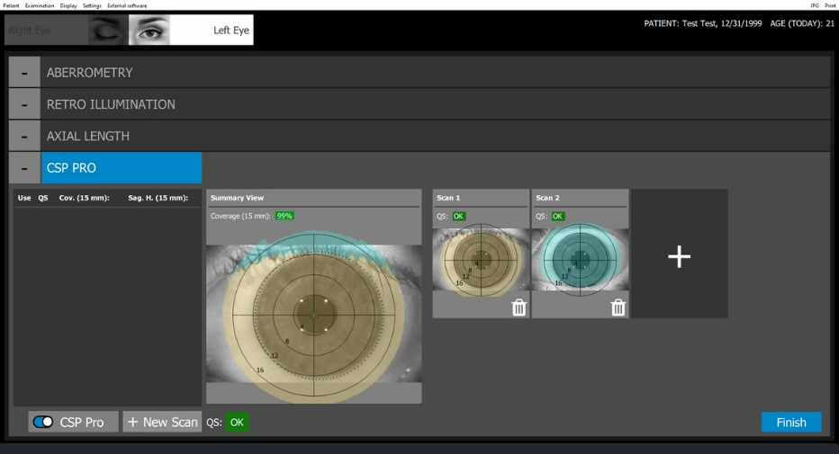
CSP Report Pro: Single-Shot Topography for Efficient & Convenient Scleral Lens Fittings
The new single-scan Scheimpflug technology of the CSP Report Pro can achieve extensive coverage in one acquisition, though practitioners can take as few or as many scans needed to capture optimal data while focusing on patient comfort. It captures topographic measurement of the cornea, sclera, and sagittal height. For patients and eye care professionals, the CSP Report Pro enables an easier process for fitting scleral and corneal lenses of every type and diameter.
The CSP Report Pro is integrated as part of the examination routine, which guides the user through the imaging process. Since neither the patient nor the device need to be repositioned, the intuitive guide enables fast examination of both eyes within just a few minutes.
Enjoy a patient-friendly imaging process since the CSP Report Pro measurement is fast, non-contact, and doesn’t require fluorescein. Practitioners also benefit because the measurement process is user-independent, reliable, straightforward, and tear film-independent.
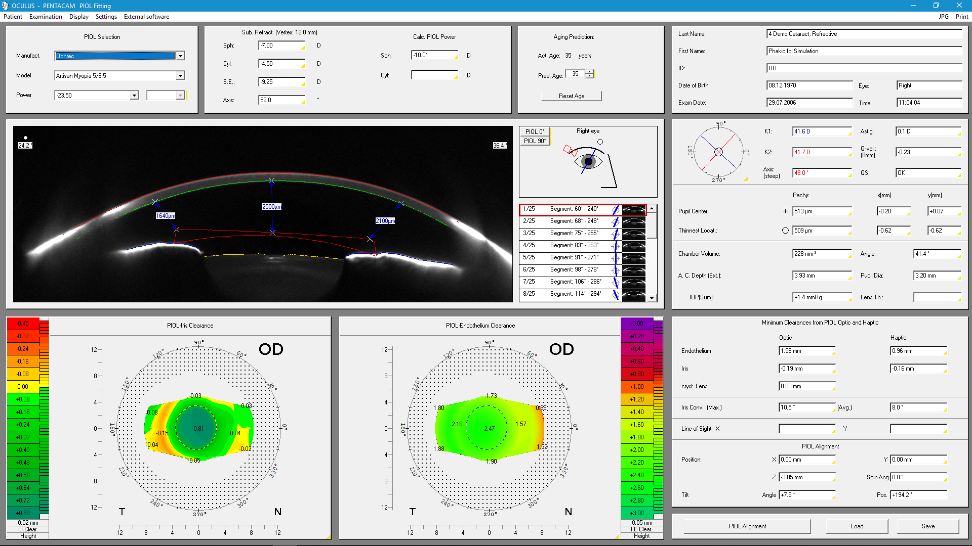
3D pIOL Simulation and Aging Prediction
Uses
- Pre-surgical planning of an iris-supported phakic anterior chamber lens
- Simulation of the post-operative position of the phakic anterior chamber lens
- Simulation of age-related lens growth and the position of the anterior chamber lens resulting from this
Details
The examiner enters the data on the patient's subjective refraction. Depending on the type of lens in question, the software calculates the necessary refractive power of the pIOL. The examiner selects a phakic IOL from the current database accordingly. The position of this pIOL in the anterior chamber is calculated in 3D and displayed in Scheimpflug images. The minimal distances between the phakic IOL and the crystalline lens as well as between the pIOL and the endothelium are calculated automatically and represented both in colour and numerically. This enables the examiner to give the patient visual information and facilitates patient selection.
- Dick et al, CATARACT & REFRACTIVE SURGERY TODAY EUROPE I JANUARY/FEBRUARY 2007