Pentacam®
The Gold Standard in anterior eye segment tomography
Since its introduction in 2002, the OCULUS Pentacam® has proven itself indispensable and has come to represent the "Gold Standard" worldwide. Here’s why: Contact-free Pentacam® measurement provides the basis for precise and reliable diagnostics and successful treatment of the anterior segment.
Comparison of Pentacam® Models
All hard- and software features at a glance
Optional Software
Pentacam® | Pentacam® HR | Pentacam® AXL Wave | |
|---|---|---|---|
| Screening Package | ◯ | ◯ | ◯ |
| Belin/Ambrósio Enhanced Ectasia Display E | ◯ | ◯ | ◯ |
| Corneal Optical Densitometry E | ◯ | ◯ | ◯ |
| Show 2 Exams | |||
| 4 Maps Selectable | |||
| Refractive Package | ◯ | ◯ | ◯ |
| Corneal Optical Densitometry E | ◯ | ◯ | ◯ |
| Refractive Display | |||
| Pachymetric Display | |||
| 4 Maps Selectable | |||
| Compare 4 Exams | |||
| 2 Exams Topometric | |||
| 2 Exams Pachymetric | |||
| Corneal Rings | |||
| Cataract Package | ◯ | ◯ | ◯ |
| PNS and 3D Cataract Analysis | |||
| Cataract Pre-OP Display | |||
| Zernike Analysis | |||
| Corneal Power Distribution | |||
| Compare 4 Exams | |||
| Show 2 Exams Topometric | |||
| Show 2 Exams Pachymetric | |||
| 4 Maps Topometric | |||
| 4 Maps Anterior Chamber | |||
| Total Corneal Refractive Power (TCRP) | |||
| True Net Power (TNP) | |||
| Anterior Chamber Depth | |||
| Anterior Chamber Angle at Scheimpflug Images | |||
| Contact Lens Fitting Package | — | ◯* | ◯ |
| CSP Report Pro / CSP Report Pro „One Shot“ E | ◯ / — | ◯ / ◯* | — / ◯ |
| Contact Lens Fitting Software incl. Fourier Analysis E | ◯ | ◯ | ◯ |
| Zernike Analysis | |||
| Compare 4 Exams | |||
| Myopia Package | — | — | ◯ |
| Growth Curves | |||
| Myopia Guide | |||
| Growth Control | |||
| GRAS Modul |
◯ optional — not available
E Module available individually
* Exclusive to Pentacam® HR units manufactured from March 2024 onwards
Standard Software & Single Licenses
| Standard Software | Pentacam® | Pentacam® HR | Pentacam® AXL Wave |
|---|---|---|---|
| Fast Screening Report | ✓ | ✓ | ✓ |
| 1 Large Color Map | ✓ | ✓ | ✓ |
| Virtual Eye | ✓ | ✓ | ✓ |
| Tomography | ✓ | ✓ | ✓ |
| 4 Maps Refractive | ✓ | ✓ | ✓ |
| General Overview | ✓ | ✓ | ✓ |
| Anterior Segment Tomography | ✓ | ✓ | ✓ |
| Topometric / KC Staging (Belin ABCD Keratoconus Staging) | ✓ | ✓ | ✓ |
| Belin ABCD Progression Display | ✓ | ✓ | ✓ |
| Iris Camera and Automatic HWTW Measurement | ✓ | ✓ | ✓ |
| 3D Anterior Chamber Analysis | ✓ | ✓ | ✓ |
| Compare 2 Exams | ✓ | ✓ | ✓ |
| Compare 2 Exams Scheimpflug Images | ✓ | ✓ | ✓ |
| Scheimpflug Image Overview | ✓ | ✓ | ✓ |
| Full Sequence Measurement | ✓ | ✓ | ✓ |
| Full Sequence Overview E | — | — | ✓ |
| Aberrometry E | — | — | ✓ |
| Iris/Retro Image | — | — | ✓ |
| Single Licenses | |||
| Holladay Report und Holladay EKR Detail Report | ◯ | ◯ | ◯ |
| 3D pIOL Simulation and Aging Prediction | — | ◯ | ◯ |
| IOL Calculator | — | ◯ | ◯ |
| DICOM | ◯ | ◯ | ◯ |
| Visual Performance | — | — | ◯ |
✓ included ◯ optional — not available
E Module available individually
All packages and single licenses are available as multiple licenses for a discounted price.
 |  | |
|---|---|---|
Scheimpflug Camera | Pentacam® | Pentacam® HR |
| Camera | digital CCD camera | digital CCD camera |
| Light source | blue LEDs (475 nm UV-free) | blue LEDs (475 nm UV-free) |
| Processor | DSP with 400m operations/s | DSP with 400m operations/s |
| Speed | 50 images in 2 seconds1) | 100 images in 2 seconds2) |
Measurement range | Pentacam® | Pentacam® HR |
| Curvature | 3 to 38 mm 9 to 99 D | 3 to 38 mm 9 to 99 D |
| Precision | ± 0.2 D | ± 0.1 D |
| Reproducibility | ± 0.2 D | ± 0.1 D |
| Operating distance | 80 mm (3.1 in) | 80 mm (3.1 in) |
Technical specifications | Pentacam® | Pentacam® HR |
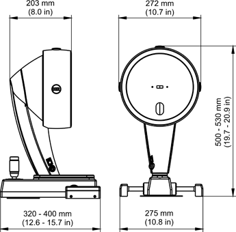
| Dimensions (W x D x H) | 275 x 320-400 x 500-530 mm (10.8 x 12.6 - 15.7 x 19.7 - 20.9 in) | 275 x 320-400 x 500-530 mm (10.8 x 12.6 - 15.7 x 19.7 - 20.9 in) |
| Weight | 10.1 kg (22.3 lbs) | 10.6 kg (23.4 lbs) |
| Power input, max. | 35 W | 42 W |
| Recommended computer specifications | CPU Intel Core i5-6600, HDD 1 TB, RAM 8 GB, MS Windows® 10 Pro, VESA, USB interface | CPU Intel Core i5-6600, HDD 1 TB, RAM 8 GB, MS Windows® 10 Pro, VESA, USB interface |
1) Scheimpflug image of the entire anterior segment
2) Cornea fine scan
The Pentacam® HR hardware differs from the one of the Pentacam® model. This means it is not possible to upgrade a Pentacam® to a Pentacam® HR. Please contact your authorized local distributor or OCULUS if you have any questions.
Technical drawing