Pentacam®
For a High-Level Entry
The Pentacam® provides you with an overall view of the anterior eye segment in a matter of seconds. Measurements are performed by auto-release and accompanied by a quality test, and are thus guaranteed to be fast, reproducible and delegable.
The Pentacam® comes with an extensive basic software package which can be extended according to your needs to include further optional software packages and modules.
Pentacam® Optional Software Modules
CSP Report
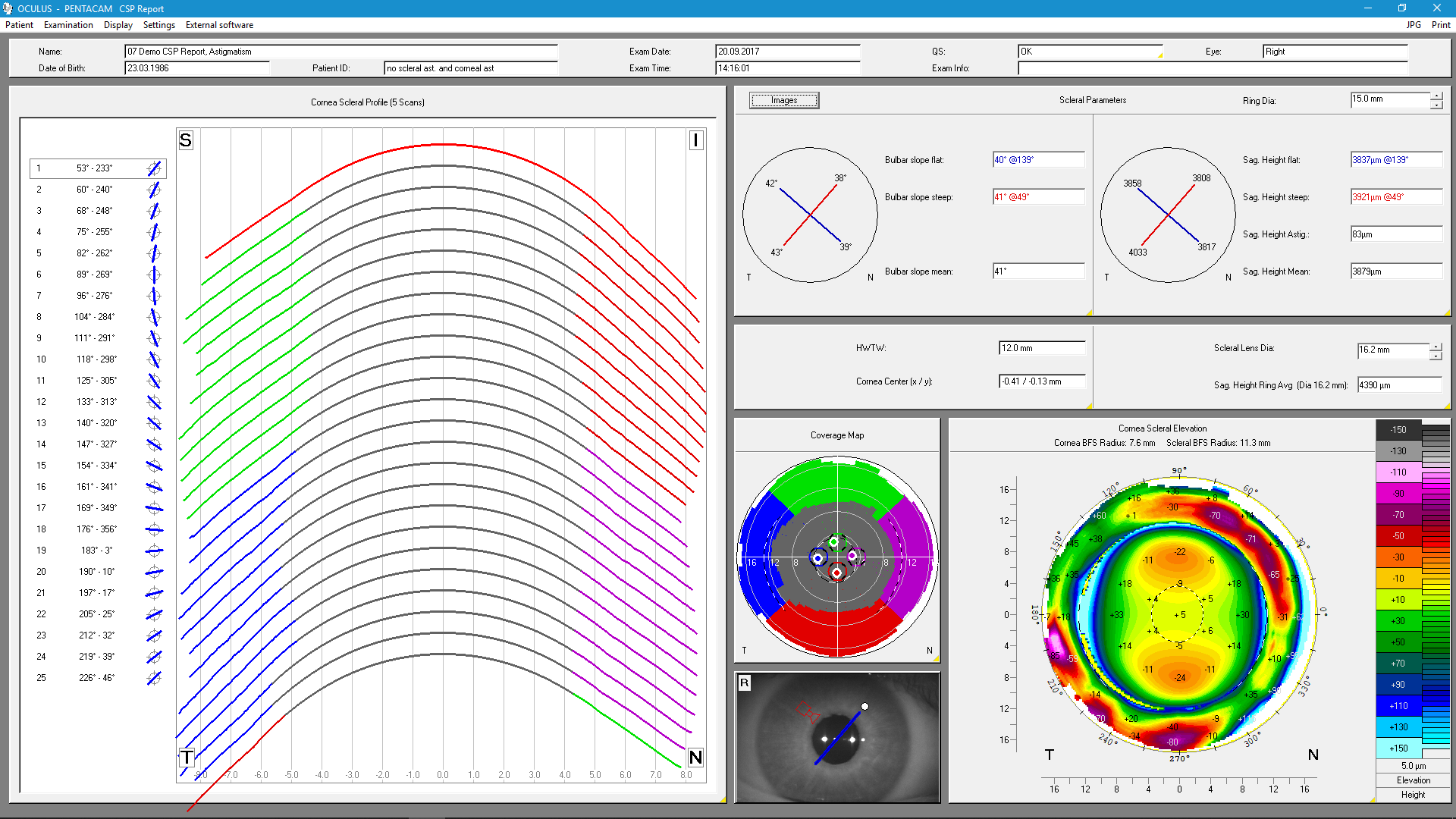
Uses
- Display of sagittal heights of cornea and sclera
- Profile of sclera for scleral lens fitting
Details
With the CSP Report, the Pentacam® now measures sagittal height, a parameter needed for scleral lens fitting. 250 Scheimpflug images covering a diameter of up to 18 mm are taken in the measuring process. All images of a Cornea Scleral Profile (CSP) scan are taken from the same visual axis without the need for eye movement. The usual Pentacam® data are recorded as well and populated into the displays already familiar to Pentacam® users. The CSP scan is a tear film independent measurement with automatic release. This means that the values from the CSP Report are as reproducible as all other data measured with the Pentacam®. A link to external fitting software for scleral lenses is available.
Refractive Package

- Free selection of reference surfaces for the colour representation of elevation data
- Corneal Optical Densitometry
- Corneal Rings
- Display of corneal thickness progression for keratoconus detection
- 4 Maps Selectable
- Show 2 Exams
- Compare 4 Exams
- Side-by-side comparison of topometric and pachymetric data
- Fourier Analysis
Cataract Package
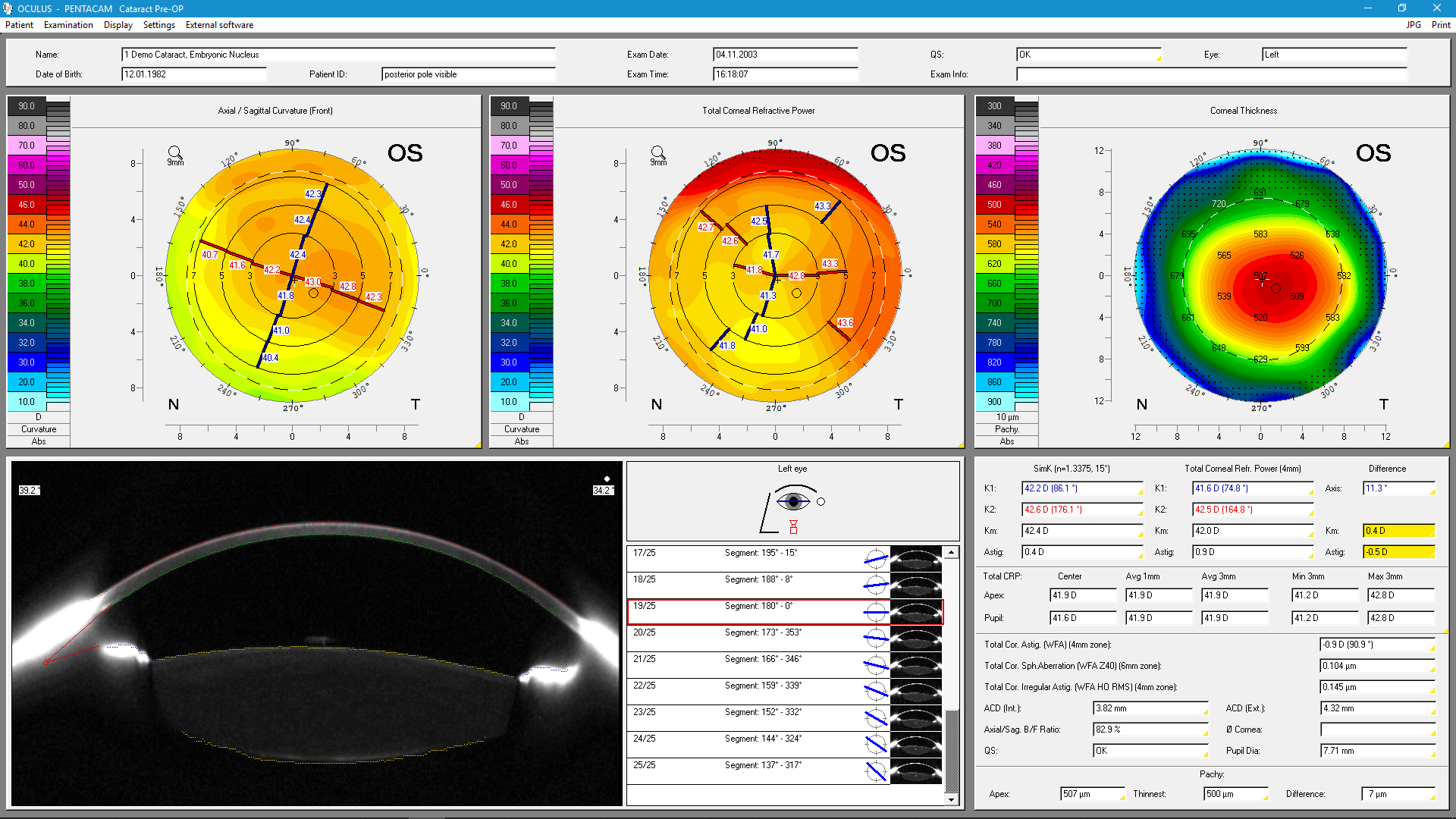

- Cataract Pre-Op Display for premium IOL selection and assessment of the optical properties of the entire cornea
- True Net Power map (TNP)
- Total Corneal Refractive Power map (TCRP)
- Corneal Power Distribution
- Zernike Analysis including normative corneal wavefront data
- PNS and 3D Cataract Analysis
- Show 2 Exams
- Compare 4 Exams
- Comparative analysis of topometric and pachymetric data
- Automatic anterior chamber angle analysis
- 4 Maps Chamber
- 4 Maps Topometric
- Anterior Chamber Depth Map
- Measurements in the Scheimpflug images
- 3D anterior chamber analysis
Corneal Optical Densitometry
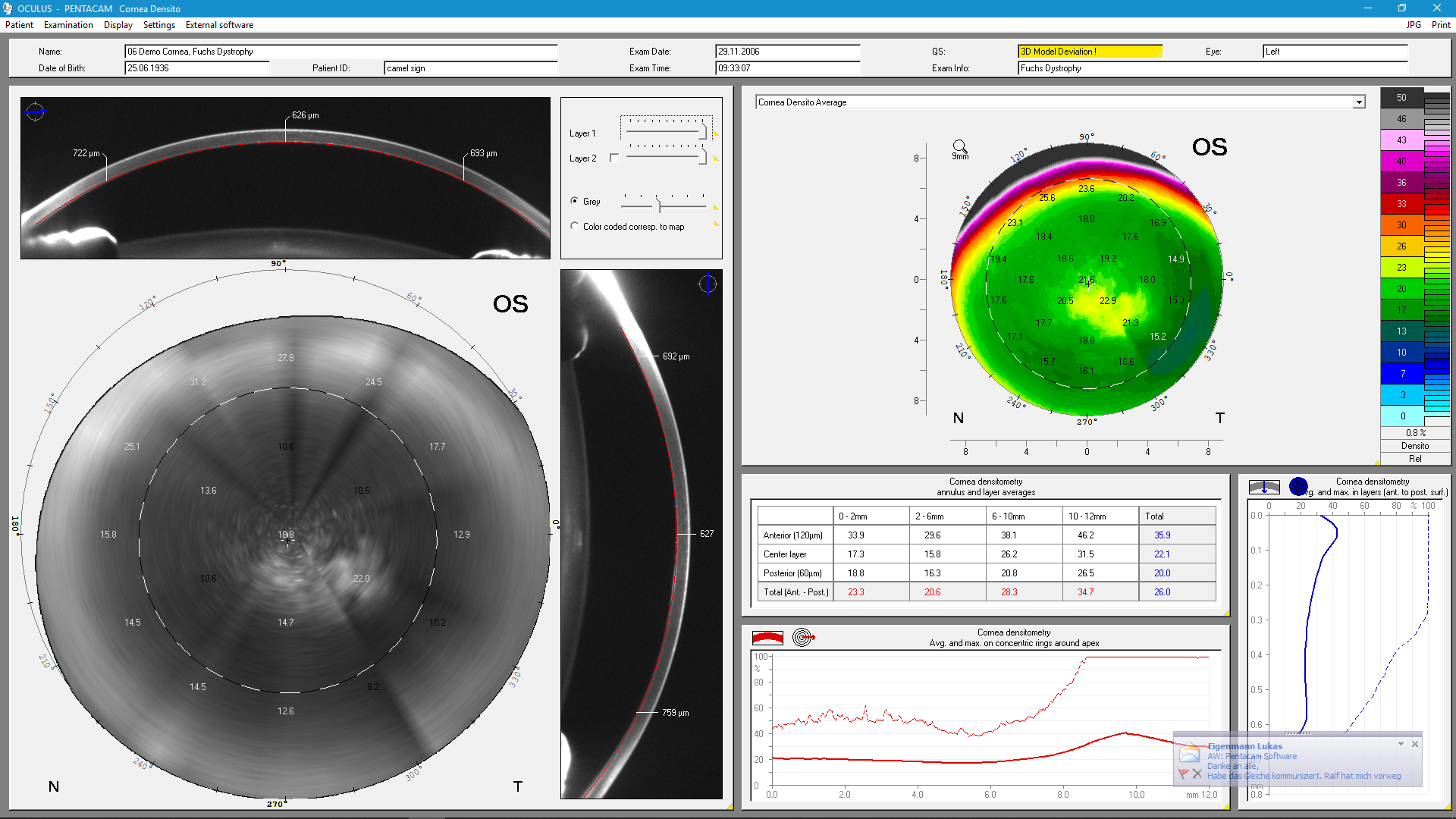
Uses
- Location of corneal opacities
- Assessment of the depth and degree of opacification
Details
This software provides a standardized method of selective quantification of corneal opacities. The maximum and average corneal opacity is displayed in a colour map. Opacities can also be viewed in selected layers. Corneal opacities are shown for defined annuli and depths, facilitating uniform evaluation. This function served as a basis in the normative data studies listed below.
- Sorcha Ni Dhubhghaill, Jos J. Rozema, Sien Jongenelen, Irene Ruiz Hidalgo, Nadia Zakaria,Marie-Jose Tassignon; Normative Values for Corneal Densitometry Analysis by Scheimpflug Optical Assessment, IOVS, January 2014, Vol. 55, No. 1, 164.
- Sorcha Ni Dhubhghaill, Jos J. Rozema, Marie-Jose Tassignon; Corneal Scheimpflug Densitometry Values measured by Pentacam in Fuchs Endothelial Dystrophy, ARVO 2014, Program Number: 2447 Poster Board Number: B0232
PNS and 3D Cataract Analysis

Uses
- Cataract classification
- Determination of lens opacities in 3D
Details
Opacities of the cornea, crystalline lens or of IOLs, e.g. PCO (posterior capsular opacification) are made visible by blue light illumination. The software allows automatic and objective three-dimensional quantification of lens opacity. This offers the advantage of an optimal follow-up of cataract patients. The Pentacam® Nucleus Staging Module, „PNS“ for short, was developed in collaboration with Dr. Donald Nixon. This parameter is helpful in optimizing the settings of phaco machines. It is also useful in femto laser assisted cataract surgery.
- Nixon et al; PREOPERATIVE CATARACT GRADING BY SCHEIMPFLUG IMAGING; doi:10.1016/j.jcrs.2009.08.032
- Meyer et al, Impact of Crystalline Lens Opacification on Effective Phacoemulsification Time in Femtosecond Laser-Assisted Cataract Surgery; AJO; doi.org/10.1016/j.ajo.2013.09.017
Holladay Report
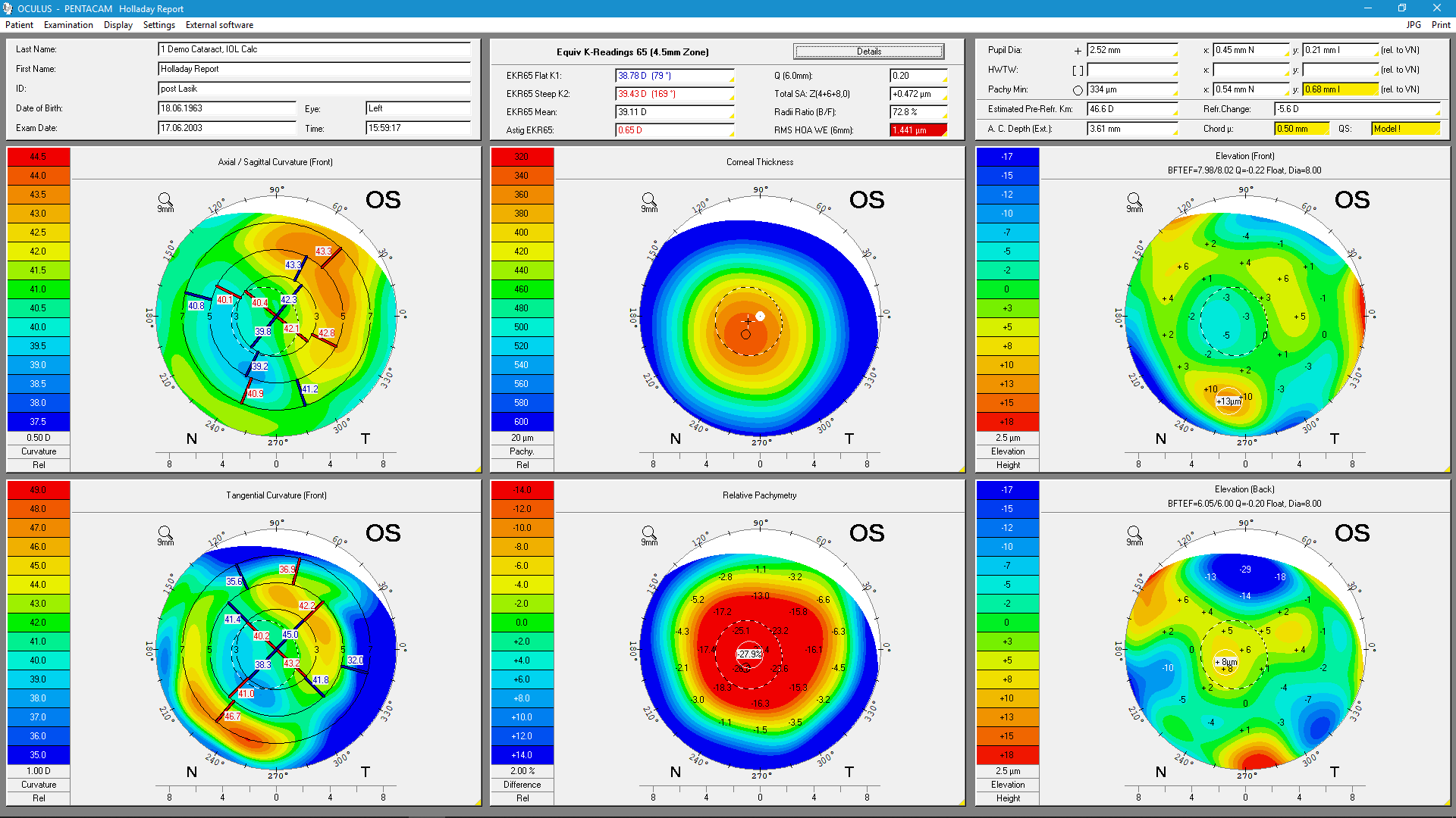
Uses
- Comprehensive comparative display of clinical data
- EKRs (equivalent keratometer readings) for IOL calculations in post-refractive patients
Details
The Holladay Report was developed in collaboration with Jack T. Holladay, M.D. It supplies data for calculating the optimal IOL refractive power for patients who have undergone refractive corneal surgery such as LASIK, PRK and RK. It determines total corneal power, displaying it in terms of EKRs for different zones.
- Holladay et al; Corneal Power Measurements Using Scheimpflug Imaging in Eyes With Prior Corneal Refractive Surgery, J Refract Surg. 2009 Oct;25(10):862-8.
- Symes et al; Automated keratometry in routine cataract surgery: Comparison of Scheimpflug and conventional values, J Cataract Refract Surg. 2011 Feb;37(2):295-301
Belin/Ambrósio Enhanced Ectasia Display
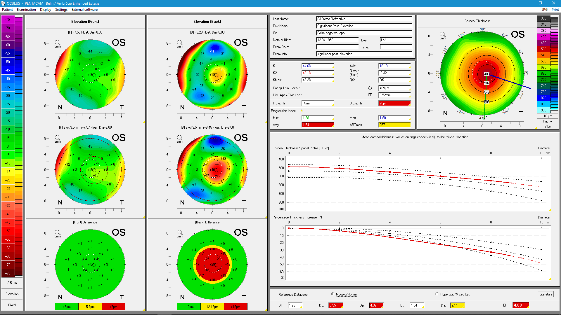
Uses
- Early ectasia detection
- Reliable detection of forme-fruste keratoconus
Details
The Belin/Ambrósio Enhanced Ectasia Display is the first screening tool to combine anterior and posterior corneal elevation data with an evaluation of corneal thickness progression in a final parameter, referred to as Index D. Besides allowing for greater overall accuracy in diagnosing ectasia it can in particular assist in the early detection of this disorder. It determines the progression of corneal thickness across an array of concentric rings centered on the thinnest point and reaching into the periphery. Two elevation maps are generated, one based on a standard reference surface and the other on an enhanced reference surface, and the differences found between them are shown in a colour code to facilitate evaluation: green for unremarkable, yellow for conspicuous, and red for potentially pathological.
- Villavicencio et al, Independent Population Validation of the Belin/Ambrosio Enhanced Ectasia Display: Implications for Keratoconus Studies and Screening, Int. Journal of Keratoconus and Ectatic Corneal Diseases, Jan-Apr 2014;3(1):1-8
- Ambrosio et al; Corneal-thickness spatial profile and corneal-volume distribution: Tomographic indices to detect keratoconus, J Cataract Refract Surg - VOL 32, NOVEMBER 2006
- Khachikian et al; Posterior Elevation in Keratoconus, Ophthalmology; Volume 116, Issue 4 , Pages 816-816.e1, April 2009
- Ambrosio et al; Corneal Ectasia After LASIK Despite Low Preoperative Risk: Tomographic and Biomechanical Findings in the Unoperated, Stable, Fellow Eye, J Refract Surg. 2010 Nov;26(11):906-11.
- Belin et al; Corneal Ectasia Risk Score: Statistical Validity and Clinical Relevance, Journal of Refractive Surgery Vol. 26, No. 4, 2010;
- Kim et al; Tomographic Normal Values for Corneal Elevation and Pachymetry in a Hyperopic Population, J Clinic Experiment Ophthalmol Volume 2, Issue 2, 1000130; ISSN:2155-9570;
- Feng et al; International values of corneal elevation in normal subjects by rotating Scheimpflug camera, J Cataract Refract Surg 2011; 37:1817-1821 Q 2011 ASCRS and ESCRS;
- Ambrosio et al; Novel Pachymetric Parameters Based on Corneal Tomography for Diagnosing Keratoconus, J Refract Surg. 2011 Oct;27(10):753-8;
- Correia et al; Topometric and Tomographic Indices for the Diagnosis of Keratoconus, International Journal of keratoconus and Eczatic Diseases, May-August 2012; 1 (2):92-99;
- Gilani et al; Comprehensive anterior segment normal values generated by rotating Scheimpflug tomography, J Cataract Refract Surg 2013; 39:1707-1712 Q 2013 ASCRS and ESCRS;
- Ambrosio et al; Corneal Ectasia After LASIK Despite Low Preoperative Risk: Tomographic and Biomechanical Findings in the Unoperated, Stable, Fellow Eye, Journal of Refractive Surgery Vol. 26, No. 11, 2010
Contact Lens Fitting incl. fourier analysis
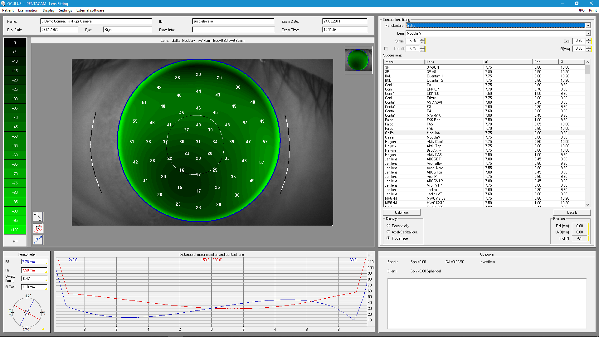
Uses
- Automatic suggestions for contact lenses
- Realistic fluo image simulation
- Integrated database containing over 845,000 lenses
Details
Dynamic fluo image simulation produces an image of how a specific contact lens from the database fits on the eye. The simulation makes it possible to adjust the inclination and position of the contact lens and includes automatic recalculation of the fluo image. The integrated and expandable database contains over 845,000 lenses. The contact lens geometries can be adjusted individually in cases where fitting is difficult. The user can establish his or her own ranking list for contact lens manufacturers.